Image of the Month
All users of the Advanced Microscopy and Imaging Center are encouraged to participate in the Image of the Month contest. To participate, simply email an image that you took with one of the Center instruments to microscopy@utk.edu with a short description of what the sample is and how you have imaged it. Only two-dimensional images will be accepted (no 3D image stacks or time-lapse sequences). At the beginning of every month, a panel of experienced microscopists will select the best image and post it on this page.
August 2018
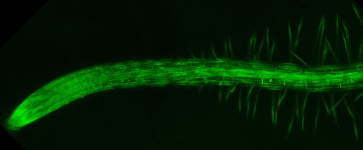
Andreas Nebenführ (Biochemistry and Cellular and Molecular Biology)
Projection of confocal images showing actin filaments in the root of Arabidopsis thaliana. These filaments form the highway system of plant cells along which organelles can be trafficked at high speeds. This is most pronounced in the root hairs that project out from the root itself.
Actin filaments were labeled with the fluorescently labeled actin-binding domain of the protein fimbrin. 31 individual confocal slices were captured in 10 µm steps at a nominal pixel size of 190 nm with the 10x/0.3 objective at a scan rate of 200 lines per second.
This image was collected on the Leica SP8X confocal microscope by Andreas Nebenführ, a faculty member in the Department of Biochemistry and Cellular and Molecular Biology.