Image of the Month
All users of the Advanced Microscopy and Imaging Center are encouraged to participate in the Image of the Month contest. To participate, simply email an image that you took with one of the Center instruments to microscopy@utk.edu with a short description of what the sample is and how you have imaged it. Only two-dimensional images will be accepted (no 3D image stacks or time-lapse sequences). At the beginning of every month, a panel of experienced microscopists will select the best image and post it on this page.
October 2018
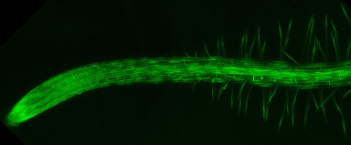
Jessica Fernandez (Biochemistry and Cellular and Molecular Biology)
Distribution of the Arabidopsis thaliana myrosinase GFP-TGG1 transiently expressed in Nicotiana benthamiana. An image stack covering 25 µm was captured with the 25x/0.9 water immersion objective on the Leica SP8X confocal microscope. Green (GFP) and red (chlorophyll) fluorescence was imaged simultaneously to show presence of introduced myrosinase and endogenous chloroplasts, respectively. The Z-stack images were maximal intensity projected into a single plane to generate this image.
Myrosinases are responsible for the hydrolysis of glucosinolates and play an important role in herbivore response. Fun fact: Glucosinolates are the compounds responsible for the pungent flavor found in mustard, cabbage and brussels sprouts!
This image was collected on the Leica SP8X confocal microscope by Jessica Fernandez, a graduate student in the lab Dr. Tessa Burch-Smith of in the Department of Biochemistry and Cellular and Molecular Biology.