Image of the Month
All users of the Advanced Microscopy and Imaging Center are encouraged to participate in the Image of the Month contest. To participate, simply email an image that you took with one of the Center instruments to microscopy@utk.edu with a short description of what the sample is and how you have imaged it. Only two-dimensional images will be accepted (no 3D image stacks or time-lapse sequences). At the beginning of every month, a panel of experienced microscopists will select the best image and post it on this page.
April 2019
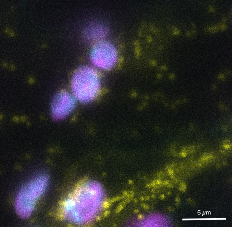
Mitochondria and chloroplasts in Nicotiana benthamiana leaf cells. Transient expression of a mitochondrial marker (yellow fluorescent protein) and a cerulean-tagged chloroplast protein (cyan) were superimposed with chlorophyll autofluorescence (shown in magenta). The different shading within the chloroplasts indicates their substructure with magenta highlighting the membrane stacks of the grana while cyan indicates specific accumulation of the protein in the stroma.
This image is a maximal intensity projection of 16 confocal images covering 6.36 µm. Cerulean and chlorophyll signals were captured simultaneously with 405 nm excitation. YFP fluorescence (514 nm excitation) was collected in a separate sequence. Images were generated on the Leica SP8 confocal microscope.
Thomas Payne is a graduate student in the lab of Andreas Nebenführ in the Department of Biochemistry & Cellular and Molecular Biology.