Image of the Month
All users of the Advanced Microscopy and Imaging Center are encouraged to participate in the Image of the Month contest. To participate, simply email an image that you took with one of the Center instruments to microscopy@utk.edu with a short description of what the sample is and how you have imaged it. Only two-dimensional images will be accepted (no 3D image stacks or time-lapse sequences). At the beginning of every month, a panel of experienced microscopists will select the best image and post it on this page.
February 2019
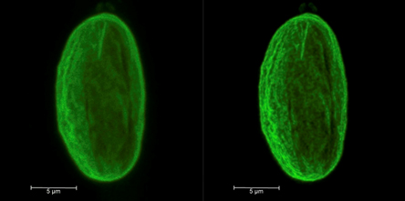
Image of the nuclear lamina from a migratory melanoma cell that has moved through a 5 µm wide constriction. The nucleus has lost its normal round shape and lamin A has irregular distribution.
Maximum intensity projection of multiple images taken at different focal planes. The left panel shows the original image while the right panel displays a deconvolved version with the new Lightning software package that is available on the Leica SP8X confocal microscope. The Lightning software improves the resolution of images by almost two-fold to reveal finer details while at the same time increasing contrast.
This image was taken by Rosela Golloshi who is a graduate student in the lab of Dr. Rachel Patton McCord in the Department of Biochemistry & Cellular and Molecular Biology.