Image of the Month
All users of the Advanced Microscopy and Imaging Center are encouraged to participate in the Image of the Month contest. To participate, simply email an image that you took with one of the Center instruments to microscopy@utk.edu with a short description of what the sample is and how you have imaged it. Only two-dimensional images will be accepted (no 3D image stacks or time-lapse sequences). At the beginning of every month, a panel of experienced microscopists will select the best image and post it on this page.
May 2018
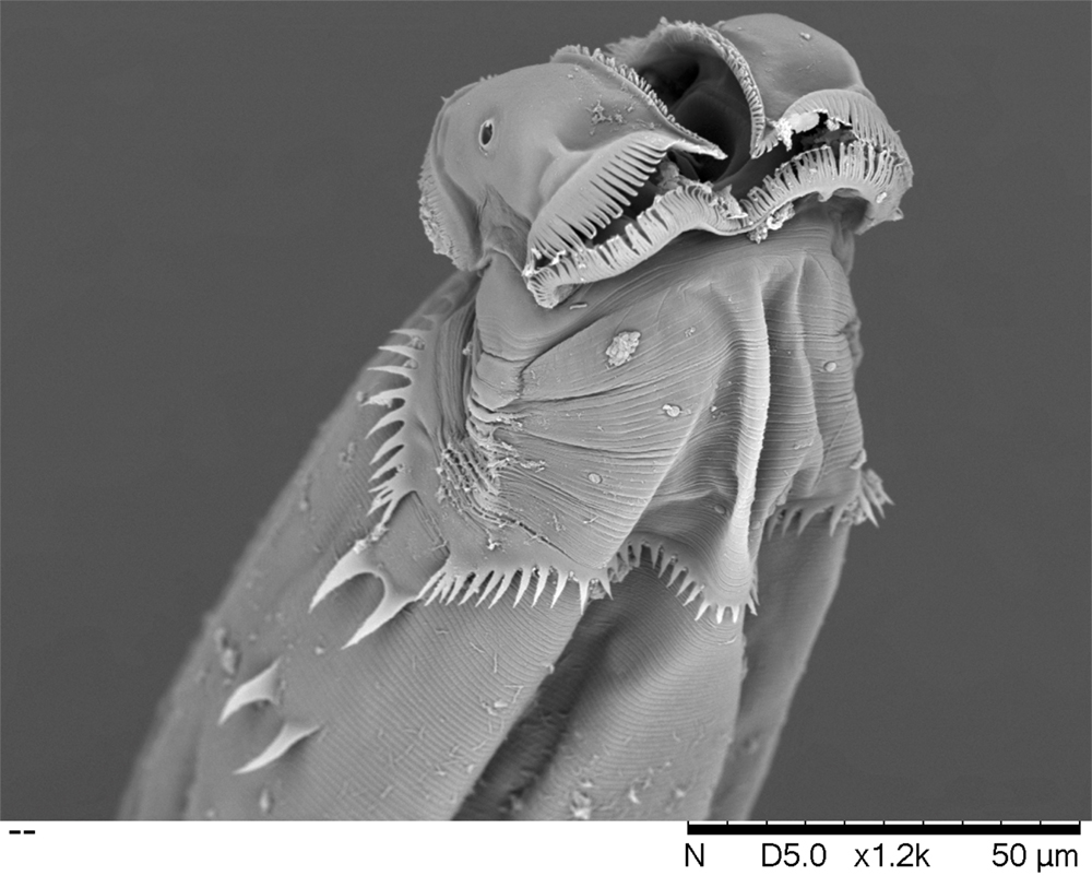
Gary Phillips, Entomology and Plant Pathology
Scanning electron micrograph of Heth pivari, a new species of nematode (roundworm) that inhabits the intestine of an indigenous millipede, Narceus gordanus, from the Ocala National Forest in Florida. The nematode feeds on the bacteria living in the gut of the millipede. The image shows a female individual with characteristic cervical structures and lateral spines. The image was captured with the secondary electron detector on the Hitachi TM3030 scanning electron microscope.